NVIDIA’s strategic approach to advancing Generative AI in Europe, enhancing AI adoption and business models….
Navigating AI Deployment: Avoiding Pitfalls and Ensuring Success
The path to AI isn’t a sprint – it’s a marathon, and businesses need to pace themselves accordingly. Those who run before they have learned to walk will falter, joining the graveyard of businesses who tried to move too quickly to reach some kind of AI…
Demed L’Her, CTO at DigitalRoute – Interview Series
Demed L’Her serves as the CTO at DigitalRoute and is a software executive with a proven track record in enterprise software strategy. He combines a strong academic background with a pragmatic approach to leadership and technology. DigitalRoute offers a portfolio designed specifically to convert raw usage…
How Combining RAG with Streaming Databases Can Transform Real-Time Data Interaction
While large language models (LLMs) like GPT-3 and Llama are impressive in their capabilities, they often need more information and more access to domain-specific data. Retrieval-augmented generation (RAG) solves these challenges by combining LLMs with information retrieval. This integration allows for smooth interactions with real-time data…
MIT releases financials and endowment figures for 2024
The Massachusetts Institute of Technology Investment Management Company (MITIMCo) announced today that MIT’s unitized pool of endowment and other MIT funds generated an investment return of 8.9 percent during the fiscal year ending June 30, 2024, as measured using valuations received within one month of fiscal year end. At the end of the fiscal year, MIT’s endowment funds totaled $24.6 billion, excluding pledges. Over the 10 years ending June 30, 2024, MIT generated an annualized return of 10.5 percent.
MIT’s endowment is intended to support current and future generations of MIT scholars with the resources needed to advance knowledge, research, and innovation. As such, endowment funds are used for Institute activities including education, research, campus renewal, faculty work, and student financial aid.
The Institute’s need-blind undergraduate admissions policy ensures that an MIT education is accessible to all qualified candidates regardless of financial resources. MIT works closely with all families who qualify for financial aid to develop an individual affordability plan tailored to their financial circumstances. In 2023-24, the average need-based MIT scholarship was $59,510. Fifty-eight percent of MIT undergraduates received need-based financial aid, and 39 percent of MIT undergraduate students received scholarship funding from MIT and other sources sufficient to cover the total cost of tuition.
Effective in fiscal 2023, MIT enhanced undergraduate financial aid, ensuring that all families with incomes below $140,000 and typical assets have tuition fully covered by scholarships. MIT further enhanced undergraduate financial aid effective in fiscal 2025, and families with incomes below $75,000 and typical assets have no expectation of parental contribution. Eighty-seven percent of seniors who graduated in academic year 2024 graduated with no debt.
MITIMCo is a unit of MIT, created to manage and oversee the investment of the Institute’s endowment, retirement, and operating funds.
MIT’s Report of the Treasurer for fiscal year 2024 was made available publicly today.
UNLV Sets the Standard for Classroom Capture with Epiphan Pearl Nexus – Videoguys
The University of Nevada, Las Vegas (UNLV) has set a new standard in educational technology by implementing Pearl Nexus for classroom capture. As a trailblazer in leveraging technology to enhance learning, UNLV has always prioritized student and faculty engagement. The university’s forward-thinking approach led them to adopt the Pearl Nexus system, an enterprise-grade solution designed to meet the evolving needs of modern education.

UNLV’s Classroom Capture Challenge
In 2018, UNLV faced a significant challenge with its existing lecture capture hardware. The system was unreliable, outdated, and not fully integrated with their AV infrastructure. With growing student and faculty needs, the university required a scalable solution that could seamlessly integrate with Panopto for content management and Crestron for room integration. As Frank Alaimo, Classroom Technology Services Manager at UNLV, explains, “our previous hardware appliances just weren’t up to the task.” The school needed an innovative solution to improve reliability and minimize maintenance.
The Pearl Nexus Solution for Classroom Capture
After extensive research, UNLV selected Pearl Nexus™ by Epiphan. The Pearl Nexus system provided everything they needed: easy integration with existing AV infrastructure, remote management through Epiphan Edge™, and fast installation. UNLV deployed over 200 Pearl Nexus units in classrooms and research spaces, each set up in less than 10 minutes. The seamless integration with Panopto and Crestron created a user-friendly experience for both faculty and students.
With Pearl Nexus, classroom capture at UNLV became streamlined and efficient. Faculty no longer needed to interact heavily with the technology, allowing them to focus on teaching, while students benefitted from having recorded lectures available for review, improving engagement and retention.

Improved Engagement and Efficiency with Pearl Nexus
Since implementing Pearl Nexus, the university has seen higher levels of student engagement. According to UNLV student Safiyaa Bintali, the ability to revisit recorded lectures has had a “positive effect” on both her learning and grades. Faculty members now direct students to lecture recordings for review, freeing up their time to address more complex questions during office hours.
The AV team at UNLV has also experienced fewer maintenance issues, allowing them to focus on strategic projects. As Frank Alaimo notes, “I’m a happy man when my phone’s not ringing,” as the reliable Pearl Nexus system allows the team to concentrate on future initiatives rather than troubleshooting old hardware.

UNLV Sets the Standard for Lecture Capture
UNLV became the first university to fully standardize on Pearl Nexus, revolutionizing how they manage classroom capture across the campus. This partnership with Epiphan has positioned UNLV as a leader in educational technology, paving the way for future innovations. Pearl Nexus has become the backbone of the university’s AV infrastructure, supporting its mission to provide students with an exceptional learning experience.
In summary, UNLV’s adoption of Pearl Nexus for classroom capture highlights the university’s commitment to leveraging cutting-edge technology. By improving student engagement, streamlining faculty workflows, and reducing AV maintenance, Pearl Nexus has set a new benchmark for lecture capture solutions in higher education. UNLV’s forward-thinking approach continues to make them a leader in delivering innovative education technology solutions.
Read the full Case Study from Epiphan HERE
Vizrt TriCaster TC1 Sucess Story – Videoguys

In this article by Jonathan Hooton, the University of Sheffield graduation ceremony is showcased as a major event for graduates and their families worldwide. To enhance the experience and share it with a global audience, the University of Sheffield uses an advanced production setup featuring the Vizrt TriCaster TC1 and NDI technology.
Before adopting the TriCaster TC1, setting up for graduation ceremonies at the University of Sheffield was a two-day process involving a team of five. The setup was complex, with many cables and equipment that made flexibility difficult. Now, with the introduction of the TriCaster TC1 and NDI technology, the production team can take advantage of the existing building infrastructure. This innovative setup eliminates the need for tangled cables and reduces time and effort.
The TriCaster TC1 serves as the heart of the operation. It enables seamless switching between five strategically placed Panasonic AW-UE150 PTZ cameras, ensuring the online audience enjoys a clear view of the stage and each graduate. The system also allows the sharing of presentations and graphics on the big screen within the Octagon Centre, while a stage camera feed highlights every graduate as they cross the stage.
One of the key features of the updated setup is its efficiency. Most of the production equipment is housed in a pre-configured flight case, affectionately called “The Beast.” This portable setup allows the team to roll into any location, connect a few cables, and be ready in just 3-4 hours, a massive improvement from the previous two-day setup. The team has also downsized from five people to just two – one handling vision mixing and the other managing camera control.
Thanks to this streamlined approach, the University of Sheffield can now reach a broader audience. Last week alone, the live stream on the Kaltura platform recorded over 18,000 views from around the globe, with on-demand viewership continuing to rise.
By adopting innovative video production technology, the University of Sheffield ensures its graduation ceremonies are shared and celebrated by graduates and families worldwide. The combination of the TriCaster TC1, NDI technology, and efficient workflows has transformed the University’s ability to host a truly global event.
Read the full article by Jonathon Hooton HERE
Learn more about Vizrt below:
A Poisoning Attack Against 3D Gaussian Splatting
A new research collaboration between Singapore and China has proposed a method for attacking the popular synthesis method 3D Gaussian Splatting (3DGS). The attack uses crafted training images of such complexity that they are likely to overwhelm an online service that allows users to create 3DGS…
A new method makes high-resolution imaging more accessible
A classical way to image nanoscale structures in cells is with high-powered, expensive super-resolution microscopes. As an alternative, MIT researchers have developed a way to expand tissue before imaging it — a technique that allows them to achieve nanoscale resolution with a conventional light microscope.
In the newest version of this technique, the researchers have made it possible to expand tissue 20-fold in a single step. This simple, inexpensive method could pave the way for nearly any biology lab to perform nanoscale imaging.
“This democratizes imaging,” says Laura Kiessling, the Novartis Professor of Chemistry at MIT and a member of the Broad Institute of MIT and Harvard and MIT’s Koch Institute for Integrative Cancer Research. “Without this method, if you want to see things with a high resolution, you have to use very expensive microscopes. What this new technique allows you to do is see things that you couldn’t normally see with standard microscopes. It drives down the cost of imaging because you can see nanoscale things without the need for a specialized facility.”
At the resolution achieved by this technique, which is around 20 nanometers, scientists can see organelles inside cells, as well as clusters of proteins.
“Twenty-fold expansion gets you into the realm that biological molecules operate in. The building blocks of life are nanoscale things: biomolecules, genes, and gene products,” says Edward Boyden, the Y. Eva Tan Professor in Neurotechnology at MIT; a professor of biological engineering, media arts and sciences, and brain and cognitive sciences; a Howard Hughes Medical Institute investigator; and a member of MIT’s McGovern Institute for Brain Research and Koch Institute for Integrative Cancer Research.
Boyden and Kiessling are the senior authors of the new study, which appears today in Nature Methods. MIT graduate student Shiwei Wang and Tay Won Shin PhD ’23 are the lead authors of the paper.
A single expansion
Boyden’s lab invented expansion microscopy in 2015. The technique requires embedding tissue into an absorbent polymer and breaking apart the proteins that normally hold tissue together. When water is added, the gel swells and pulls biomolecules apart from each other.
The original version of this technique, which expanded tissue about fourfold, allowed researchers to obtain images with a resolution of around 70 nanometers. In 2017, Boyden’s lab modified the process to include a second expansion step, achieving an overall 20-fold expansion. This enables even higher resolution, but the process is more complicated.
“We’ve developed several 20-fold expansion technologies in the past, but they require multiple expansion steps,” Boyden says. “If you could do that amount of expansion in a single step, that could simplify things quite a bit.”
With 20-fold expansion, researchers can get down to a resolution of about 20 nanometers, using a conventional light microscope. This allows them see cell structures like microtubules and mitochondria, as well as clusters of proteins.
In the new study, the researchers set out to perform 20-fold expansion with only a single step. This meant that they had to find a gel that was both extremely absorbent and mechanically stable, so that it wouldn’t fall apart when expanded 20-fold.
To achieve that, they used a gel assembled from N,N-dimethylacrylamide (DMAA) and sodium acrylate. Unlike previous expansion gels that rely on adding another molecule to form crosslinks between the polymer strands, this gel forms crosslinks spontaneously and exhibits strong mechanical properties. Such gel components previously had been used in expansion microscopy protocols, but the resulting gels could expand only about tenfold. The MIT team optimized the gel and the polymerization process to make the gel more robust, and to allow for 20-fold expansion.
To further stabilize the gel and enhance its reproducibility, the researchers removed oxygen from the polymer solution prior to gelation, which prevents side reactions that interfere with crosslinking. This step requires running nitrogen gas through the polymer solution, which replaces most of the oxygen in the system.
Once the gel is formed, select bonds in the proteins that hold the tissue together are broken and water is added to make the gel expand. After the expansion is performed, target proteins in tissue can be labeled and imaged.
“This approach may require more sample preparation compared to other super-resolution techniques, but it’s much simpler when it comes to the actual imaging process, especially for 3D imaging,” Shin says. “We document the step-by-step protocol in the manuscript so that readers can go through it easily.”
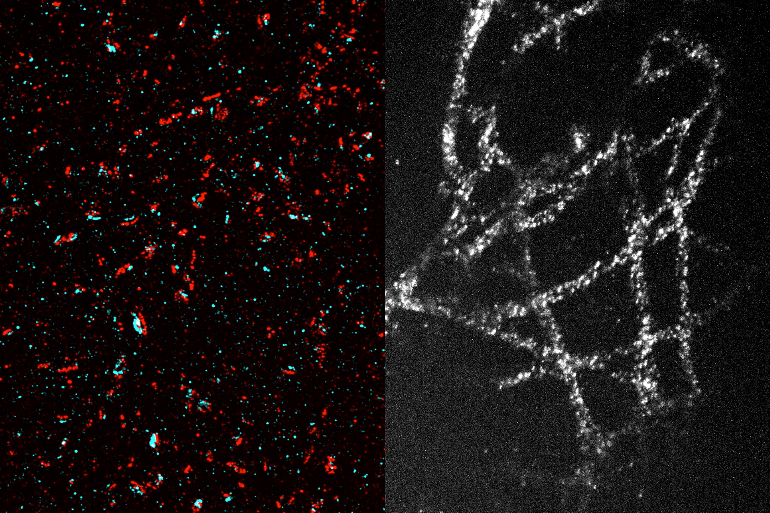
Imaging tiny structures
Using this technique, the researchers were able to image many tiny structures within brain cells, including structures called synaptic nanocolumns. These are clusters of proteins that are arranged in a specific way at neuronal synapses, allowing neurons to communicate with each other via secretion of neurotransmitters such as dopamine.
In studies of cancer cells, the researchers also imaged microtubules — hollow tubes that help give cells their structure and play important roles in cell division. They were also able to see mitochondria (organelles that generate energy) and even the organization of individual nuclear pore complexes (clusters of proteins that control access to the cell nucleus).
Wang is now using this technique to image carbohydrates known as glycans, which are found on cell surfaces and help control cells’ interactions with their environment. This method could also be used to image tumor cells, allowing scientists to glimpse how proteins are organized within those cells, much more easily than has previously been possible.
The researchers envision that any biology lab should be able to use this technique at a low cost since it relies on standard, off-the-shelf chemicals and common equipment such confocal microscopes and glove bags, which most labs already have or can easily access.
“Our hope is that with this new technology, any conventional biology lab can use this protocol with their existing microscopes, allowing them to approach resolution that can only be achieved with very specialized and costly state-of-the-art microscopes,” Wang says.
The research was funded, in part, by the U.S. National Institutes of Health, an MIT Presidential Graduate Fellowship, U.S. National Science Foundation Graduate Research Fellowship grants, Open Philanthropy, Good Ventures, the Howard Hughes Medical Institute, Lisa Yang, Ashar Aziz, and the European Research Council.
Tiny magnetic discs offer remote brain stimulation without transgenes
Novel magnetic nanodiscs could provide a much less invasive way of stimulating parts of the brain, paving the way for stimulation therapies without implants or genetic modification, MIT researchers report.
The scientists envision that the tiny discs, which are about 250 nanometers across (about 1/500 the width of a human hair), would be injected directly into the desired location in the brain. From there, they could be activated at any time simply by applying a magnetic field outside the body. The new particles could quickly find applications in biomedical research, and eventually, after sufficient testing, might be applied to clinical uses.
The development of these nanoparticles is described in the journal Nature Nanotechnology, in a paper by Polina Anikeeva, a professor in MIT’s departments of Materials Science and Engineering and Brain and Cognitive Sciences, graduate student Ye Ji Kim, and 17 others at MIT and in Germany.
Deep brain stimulation (DBS) is a common clinical procedure that uses electrodes implanted in the target brain regions to treat symptoms of neurological and psychiatric conditions such as Parkinson’s disease and obsessive-compulsive disorder. Despite its efficacy, the surgical difficulty and clinical complications associated with DBS limit the number of cases where such an invasive procedure is warranted. The new nanodiscs could provide a much more benign way of achieving the same results.
Over the past decade other implant-free methods of producing brain stimulation have been developed. However, these approaches were often limited by their spatial resolution or ability to target deep regions. For the past decade, Anikeeva’s Bioelectronics group as well as others in the field used magnetic nanomaterials to transduce remote magnetic signals into brain stimulation. However, these magnetic methods relied on genetic modifications and can’t be used in humans.
Since all nerve cells are sensitive to electrical signals, Kim, a graduate student in Anikeeva’s group, hypothesized that a magnetoelectric nanomaterial that can efficiently convert magnetization into electrical potential could offer a path toward remote magnetic brain stimulation. Creating a nanoscale magnetoelectric material was, however, a formidable challenge.
Kim synthesized novel magnetoelectric nanodiscs and collaborated with Noah Kent, a postdoc in Anikeeva’s lab with a background in physics who is a second author of the study, to understand the properties of these particles.
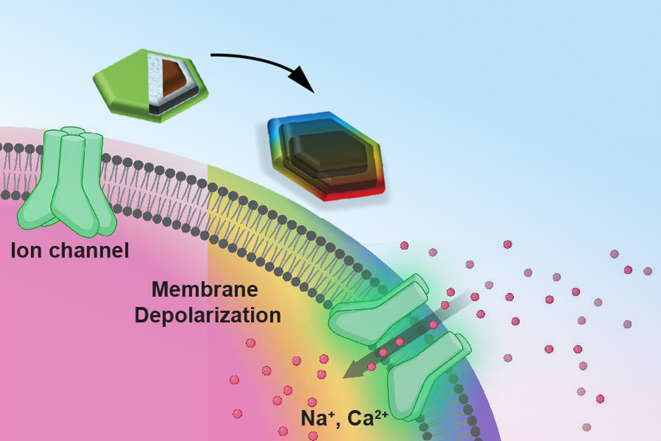
The structure of the new nanodiscs consists of a two-layer magnetic core and a piezoelectric shell. The magnetic core is magnetostrictive, which means it changes shape when magnetized. This deformation then induces strain in the piezoelectric shell which produces a varying electrical polarization. Through the combination of the two effects, these composite particles can deliver electrical pulses to neurons when exposed to magnetic fields.
One key to the discs’ effectiveness is their disc shape. Previous attempts to use magnetic nanoparticles had used spherical particles, but the magnetoelectric effect was very weak, says Kim. This anisotropy enhances magnetostriction by over a 1000-fold, adds Kent.
The team first added their nanodiscs to cultured neurons, which allowed then to activate these cells on demand with short pulses of magnetic field. This stimulation did not require any genetic modification.
They then injected small droplets of magnetoelectric nanodiscs solution into specific regions of the brains of mice. Then, simply turning on a relatively weak electromagnet nearby triggered the particles to release a tiny jolt of electricity in that brain region. The stimulation could be switched on and off remotely by the switching of the electromagnet. That electrical stimulation “had an impact on neuron activity and on behavior,” Kim says.
The team found that the magnetoelectric nanodiscs could stimulate a deep brain region, the ventral tegmental area, that is associated with feelings of reward.
The team also stimulated another brain area, the subthalamic nucleus, associated with motor control. “This is the region where electrodes typically get implanted to manage Parkinson’s disease,” Kim explains. The researchers were able to successfully demonstrate the modulation of motor control through the particles. Specifically, by injecting nanodiscs only in one hemisphere, the researchers could induce rotations in healthy mice by applying magnetic field.
The nanodiscs could trigger the neuronal activity comparable with conventional implanted electrodes delivering mild electrical stimulation. The authors achieved subsecond temporal precision for neural stimulation with their method yet observed significantly reduced foreign body responses as compared to the electrodes, potentially allowing for even safer deep brain stimulation.
The multilayered chemical composition and physical shape and size of the new multilayered nanodiscs is what made precise stimulation possible.
While the researchers successfully increased the magnetostrictive effect, the second part of the process, converting the magnetic effect into an electrical output, still needs more work, Anikeeva says. While the magnetic response was a thousand times greater, the conversion to an electric impulse was only four times greater than with conventional spherical particles.
“This massive enhancement of a thousand times didn’t completely translate into the magnetoelectric enhancement,” says Kim. “That’s where a lot of the future work will be focused, on making sure that the thousand times amplification in magnetostriction can be converted into a thousand times amplification in the magnetoelectric coupling.”
What the team found, in terms of the way the particles’ shapes affects their magnetostriction, was quite unexpected. “It’s kind of a new thing that just appeared when we tried to figure out why these particles worked so well,” says Kent.
Anikeeva adds: “Yes, it’s a record-breaking particle, but it’s not as record-breaking as it could be.” That remains a topic for further work, but the team has ideas about how to make further progress.
While these nanodiscs could in principle already be applied to basic research using animal models, to translate them to clinical use in humans would require several more steps, including large-scale safety studies, “which is something academic researchers are not necessarily most well-positioned to do,” Anikeeva says. “When we find that these particles are really useful in a particular clinical context, then we imagine that there will be a pathway for them to undergo more rigorous large animal safety studies.”
The team included researchers affiliated with MIT’s departments of Materials Science and Engineering, Electrical Engineering and Computer Science, Chemistry, and Brain and Cognitive Sciences; the Research Laboratory of Electronics; the McGovern Institute for Brain Research; and the Koch Institute for Integrative Cancer Research; and from the Friedrich-Alexander University of Erlangen, Germany. The work was supported, in part, by the National Institutes of Health, the National Center for Complementary and Integrative Health, the National Institute for Neurological Disorders and Stroke, the McGovern Institute for Brain Research, and the K. Lisa Yang and Hock E. Tan Center for Molecular Therapeutics in Neuroscience.