President Biden has issued the US’ first-ever National Security Memorandum (NSM) on AI, addressing how the nation approaches the technology from a security perspective. The memorandum, which builds upon Biden’s earlier executive order on AI, is founded on the premise that cutting-edge AI developments will substantially…
NETGEAR Showcases Enterprise-Class M4350 Series AV Switches at IBC 202 – Videoguys

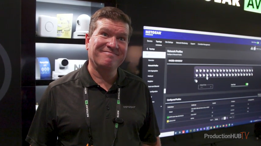
At the IBC 2024 Show in Amsterdam, NETGEAR unveiled its Enterprise-Class M4350 Series AV Switches, specifically designed to meet the demands of AV over IP (AVoIP) networks. These advanced AV switches are ideal for businesses seeking reliable, high-performance solutions for their audio, video, and lighting setups. The M4350 series stands out for its enterprise-class hardware, redundant power supplies, and support for 100G uplinks, ensuring top-tier performance for AVoIP applications.
One of the key highlights of the M4350 switches is the NETGEAR AV user interface and the Engage Controller, both equipped with pre-configured profiles to support major audio, video, and lighting protocols. The switches also feature IGMP Plus™ with Auto-LAG and Auto-Trunk, offering seamless multicast installation. Additional features like advanced IPv4/IPv6 security, intelligent thermal and acoustic controls, and PoE+ and Ultra90 PoE++ support for up to 90W per device make these switches a versatile choice for a wide range of AV installations.
With over 200 switching products in its portfolio, NETGEAR offers flexible options for AV over IP solutions, supporting speeds from 1Gbps to 100Gbps per port. This extensive product line can also power PoE cameras and security devices, providing a scalable and customizable network setup. NETGEAR’s switches can be tailored to meet specific customer needs, with room for future expansion, ensuring businesses can grow their AV infrastructure without limitations.
As video over Ethernet continues to rise in popularity, NETGEAR’s M4350 series meets the growing demand with products designed for ProAV installations. Whether you need modular switches for copper, fiber, or HDMI inputs, or a solution that fits a specific budget, NETGEAR has the expertise to guide businesses toward the perfect AV solution. These AVoIP switches offer flexible mounting options and can be deployed in a variety of environments, from university campuses to live event productions.
For businesses looking to upgrade their AV infrastructure, NETGEAR’s M4350 Series offers the perfect combination of performance, flexibility, and scalability to meet the evolving demands of AV over IP networks.
Read the full article from ProductionHub HERE
Learn more about NETGEAR below:
Building the Ultimate Sports Production System with PTZOptics, vMix, a – Videoguys
When it comes to sports production, having the right tools and technology can transform any game into a professional-grade broadcast. Whether you’re covering high school football, basketball tournaments, or college soccer, the ability to deliver high-quality live streams and multi-camera productions is becoming more important than ever. Fortunately, creating a powerful sports production system has never been easier thanks to the innovative products available from PTZOptics, vMix, and more. In this blog post, we’ll explore the key components needed to build your ultimate sports production setup and how these products work together to enhance your live broadcasts.

PTZOptics Cameras: Versatile and Dynamic for Every Sport
The backbone of any sports production system is the camera, and PTZOptics offers some of the best PTZ (pan-tilt-zoom) cameras on the market for capturing live sports. These cameras are incredibly versatile, making them ideal for covering everything from wide-field shots to up-close action. With motorized pan, tilt, and zoom capabilities, PTZ cameras can be remotely controlled, allowing a single operator to manage multiple cameras during a live broadcast.
Why PTZOptics?
- Remote Control: PTZOptics cameras can be controlled via IP, serial, or even wirelessly using PTZOptics’ Wireless Cable for more flexibility in camera placement.
- NDI Integration: These cameras are NDI-enabled, meaning they can be easily integrated into any IP-based production workflow, sending high-quality video feeds over a network with low latency.
- 4K and 1080p Resolution Options: Depending on your production needs, PTZOptics cameras offer both 4K and 1080p resolution models, ensuring you capture every detail of the action on the field or court.
PTZOptics cameras are also a fantastic option for student-run production programs, providing hands-on experience with professional equipment while being user-friendly enough for beginners.

vMix: The Heart of Your Production Workflow
At the center of your sports production system is vMix, a live video production software that gives you all the tools needed to create a professional broadcast. vMix supports multiple camera inputs, live switching, graphics, instant replay, and more, all from one powerful platform. Whether you’re running a simple two-camera setup or a complex multi-camera production with overlays and replays, vMix makes it easy to manage your broadcast with just a few clicks.
Why Choose vMix?
- Multi-Camera Input Support: vMix allows you to bring in multiple video sources from PTZOptics cameras, webcams, remote contributors via Zoom, and even mobile devices.
- Instant Replay: Perfect for sports production, vMix’s instant replay feature lets you capture and replay key moments in real time, adding a professional touch to your live streams.
- NDI Support: vMix works seamlessly with NDI, allowing you to bring in video feeds from your PTZOptics cameras, video switchers, and other IP-connected devices with minimal setup.
- Remote Control and Web Interface: vMix can be controlled remotely through its built-in web interface or integrated with Elgato StreamDeck for one-touch control of transitions, replays, and more.
For sports broadcasters, vMix is a must-have, offering a comprehensive production environment that scales with your needs, whether you’re covering a single game or multiple events simultaneously.

SuperJoy Joystick: Precision Camera Control
For those looking for tactile, real-time control over PTZ cameras during a live broadcast, the PTZOptics SuperJoy Joystick is a perfect addition to your sports production system. This intuitive controller allows you to manage multiple PTZ cameras from a central location with smooth, precise camera movements.
SuperJoy Key Features:
- IP and Serial Control: The SuperJoy can control PTZOptics cameras over IP or through serial connections, giving you flexibility in how you set up your production system.
- Pre-Programmed Presets: Save camera positions as presets, allowing you to quickly switch between wide shots, close-ups, and other camera angles at the push of a button.
- HDMI Output: Monitor your camera feeds in real-time through the SuperJoy’s HDMI output, which can connect to an external display to view the camera’s field of view as you operate.
For sports productions, where quick and accurate camera movements are crucial to capturing the action, the SuperJoy adds the hands-on precision you need to ensure your broadcast is smooth and professional.
Case Study: Salesianum School Elevates Sports Broadcasting with PTZOptics and vMix
[embedded content]
One example of how PTZOptics cameras and vMix software can transform a sports production system is the recent setup at Salesianum School in Wilmington, Delaware. The school has implemented a multi-camera sports production system featuring six PTZOptics cameras, all controlled through the SuperJoy joystick and integrated with vMix for live streaming, instant replays, and real-time graphics overlays.
Using PTZOptics Hive, the school’s production team can remotely control cameras across multiple sports venues, allowing them to broadcast football, soccer, and basketball games with ease. The system is run by student operators, providing them with hands-on experience in live video production while delivering professional-quality streams for fans, alumni, and families.
The flexibility and scalability of this system have made it a game-changer for Salesianum, allowing them to produce high-quality broadcasts across different sports with a minimal crew. The integration of PTZOptics cameras and vMix has streamlined their workflow and made it easier to capture every play, ensuring no moment is missed.

Hive: Cloud-Based Remote Production
For productions that require remote control and management of video feeds from multiple locations, PTZOptics Hive provides a cloud-based solution. Hive allows you to control PTZOptics cameras, manage video streams, and monitor camera feeds all through a single interface, regardless of where your event is happening.
What Hive Brings to Sports Production:
- Remote Camera Control: Perfect for schools or sports organizations with multiple fields or venues, Hive lets you manage all cameras remotely through the cloud.
- Scalability: Hive can handle large-scale productions, making it easy to control and produce games happening in different locations without needing to be on-site.
- NDI Integration: Just like PTZOptics cameras and vMix, Hive uses NDI for video feeds, making it simple to integrate into your production workflow.
Hive is a great tool for larger sports organizations or schools that want to centralize their production efforts across multiple venues, making it easier to manage camera systems and streamline live broadcasts.
Tying It All Together
Building a sports production system doesn’t have to be complicated. With PTZOptics cameras, vMix production software, the SuperJoy joystick, and Hive cloud-based management, you can create a robust, scalable system that delivers professional-quality live sports broadcasts. Whether you’re covering high school athletics or semi-pro leagues, these products are designed to work seamlessly together, ensuring your production workflow is smooth and efficient.
For schools, universities, or any organization looking to elevate their sports broadcasts, this combination of tools is perfect for creating high-quality content that engages fans, alumni, and families. And with the added benefit of remote production capabilities, your system can grow as your needs evolve.
Learn more about PTZOptics below:
AI is helping brands avoid controversial influencer partnerships – AI News
Influencer partnerships can be great for brands looking to pump out content that promotes their products and services in an authentic way. These types of engagements can yield significant brand awareness and brand sentiment lift, but they can be risky too. Social media stars are unpredictable…
Intern allegedly sabotages ByteDance AI project, leading to dismissal
ByteDance, the creator of TikTok, recently experienced a security breach involving an intern who allegedly sabotaged AI model training. The incident, reported on WeChat, raised concerns about the company’s security protocols in its AI department. In response, ByteDance clarified that while the intern disrupted AI commercialisation…
The Friday Roundup – Speed Ramping and Masking Basics
Video Editing Speed Ramping Most video editing software these days allows the ability to “speed ramp” all or sections of a video clip. To clarify, speed ramping is a term used to describe speeding up or slowing down a part of a clip to achieve an…
Augment Code Launches AI-Powered Platform to Revolutionize Team-Based Software Development
In a major move set to redefine the future of software development, Augment Code has emerged from stealth mode to unveil the first AI-powered coding platform designed specifically for development teams. With its proprietary AI technology, the platform offers an unprecedented level of contextual understanding, making…
The New Proactive CX: Generative AI Meets Customer Service
Generative AI (GenAI) is reshaping customer engagement in ways previously unimaginable. While it’s still early in its adoption, measurable business results are already being seen. According to a study by McKinsey, AI-driven customer engagement strategies have the potential to increase business revenues by up to 30%…
Dr. Ron Boucher, Chief Medical Officer of Teleradiology at Experity – Interview Series
Dr. Ron Boucher serves as the Chief Medical Officer of Teleradiology at Experity, a software and services company focused on the U.S. urgent care market. Experity offers an integrated operating system that includes electronic medical records, practice management, patient engagement, billing, teleradiology, business intelligence, and consulting…
A Game-Changer for AI: The Tsetlin Machine’s Role in Reducing Energy Consumption
The rapid rise of Artificial Intelligence (AI) has transformed numerous sectors, from healthcare and finance to energy management and beyond. However, this growth in AI adoption has resulted in a significant issue of energy consumption. Modern AI models, particularly those based on deep learning and neural…

